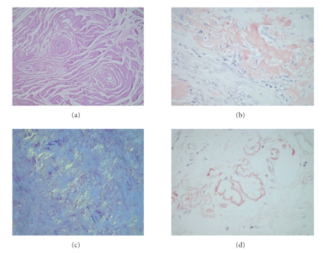
Figure 3.
(a) Closer view of amorphous, fibrillary matrix deposition (hematoxylin and eosin, ×20). (b) Peach-red staining of the material in Congo-red staining (Congo-red, ×20). (c) Apple-green birefringence under polarized light (Congo-red, polarization microscope, ×20). (d) Amyloid deposition in vessel walls (Congo-red, ×20).

