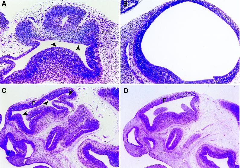
Figure 4.
Neuroepithelial abnormalities in mutant embryos. (A) Forebrain of an E10.5 Tsc2Ek/Ek mutant embryo. (B) Forebrain of wild-type embryo at E10.5, which is more rounded and inflated than the forebrain of mutant embryo shown in A. (C) Head of Tsc2Ek/Ek embryo at E11.5 with papillary overgrowth of midbrain neuroepithelium, indicated by arrows. Forebrain is misshapen and disorganized. (D) Head of wild-type embryo at E11.5. (A and C) Arrows indicate areas of thickening and folding of neuroepithelium in mutant embryos. F, forebrain.

