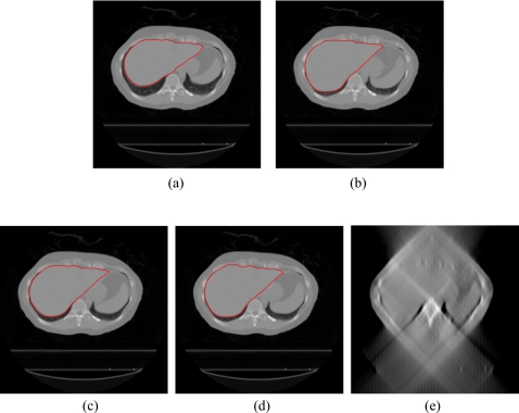
Figure 4.
DTS reconstruction of liver patient data using prior based and FBP based methods with 60°–90° scan angles. (The contours of the liver are shown in the CBCT and prior based DTS images.) (a) The prior CBCT image CBCTprior, (b) the new CBCT image CBCTnew, (c) prior based 60° DTSnew, (d) prior based 90° DTSnew, (e) FBP based 90° DTSnew.

