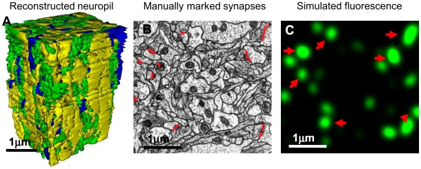
Figure 1. Electron Microscopy reconstruction data.
A) Using a 130 µm3 block of hippocampal neuropil imaged with high-resolution electron microscopy, we investigate the possibility of detecting individual synapses optically. This block was fully segmented by the author, and all synapses were explicitly labeled. For illustration purposes, here is shown the 3D model of the reconstruction of all neuronal processes in said block, colored according to process type – yellow for dendrites, green for axons, and blue for glial processes. B) An example of the manual markup of the synapses within one electron micrograph (red lines). C) Simulated fluorescence from marked synapses (here, for isotropic diffraction limited microscopy, IDLM). Red arrows indicate synapses located directly inside shown EM section (B).

