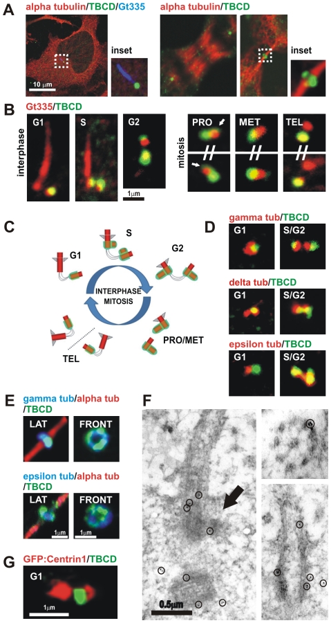
Figure 1. TBCD is concentrated in centrioles and midbodies.
(A) Confocal-microscopic images of a HeLa cell (ATCCR number: CCL-2™) immunostained for tubulin, TBCD, and GT335 (which labels glutamylated tubulin at the centrioles and primary cilium), where a single spot of TBCD is observed close to the basal body of the cilium. HeLa cells in anaphase and undergoing cytokinesis display clear centrosomal labelling and recruitment of TBCD to the midbody ring, respectively. (B) High-resolution confocal-microscopic section images of doubly immunostained centrosomes of HeLa cells. The cells were photographed at different stages of the cell cycle, labelled with GT335 and anti-TBCD antibody. This tubulin cofactor is observed at one of the centrioles during G1. Two stronger additional TBCD signals immediately adjacent to the existing centrioles, devoid of GT335 staining, were stained at G1. By late S three TBCD spots were detected. By prophase, most dividing cells contained two strong TBCD spots, one in each centrosome. Partial TBCD halos surrounding older centrioles were also observed (arrows). At the end of telophase, only one of the centrioles in each daughter cell was labelled for TBCD. (C) Diagram of the distribution of TBCD during the centriolar cycle; centrioles, procentrioles, and primary cilium are shown in red and TBCD is shown in green. (D) Partial co-localization of the TBCD signal with γ- and δ-tubulins was observed throughout the cell cycle. The ε-tubulin signal concentrated at the mother centriole during G1 did not co-localize with TBCD. Later in the cell cycle, both proteins appeared to partially co-localize. (E) TBCD accumulated at the midbody, where γ- and ε-tubulins were also detected. Lateral and frontal views of the structure correspond to different cells. (F) Immuno-electron-microscopic analysis of TBCD on murine epithelial cells. (Left) TBCD localises on the proximal region of the basal body (the original mother centriole) during procentriole assembly (arrow) S stage. The former daughter centriole is also observed in the section (gold particles are outlined with circles). (Right top) TBCD labelling was also detected at the proximal ends of basal bodies and (Right bottom) at the outer microtubule doublets of motile tracheal cilia. (G) Immunostaining of HeLa cells transfected with GFP-centrin1 (red, pseudocolor) confirm TBCD localization at the proximal end of the daughter centriole.

