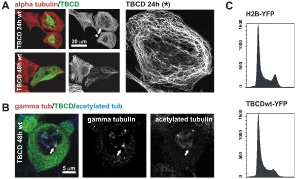
Figure 2. TBCD overexpression resulted in microtubule detachment from the centrosome and spindle abnormalities.
(A) Confocal-microscopic projected images of a HeLa cell overexpressing HsTBCD doubly immunostained for tubulin and TBCD. (Top) Microtubule release from the centrosome was clear in most cells 15 h after transfection (arrow). (Bottom) Microtubule network destruction occurred approximately 30 h after transfection. Right panel shows a closer view of the microtubule pattern of the cell labelled with an arrow.(B) TBCD overexpression also produced aberrant mitotic figures, where acentriolar supernumerary MTOCs containing γ-tubulin were observed (arrow). (C) Flow-cytometric profiles of cells (>10,000 cells per profile) labelled with Hoechst and transfected with HsTBCD–YFP or a control–YFP construct. Clear G1 arrest, typical of the loss of centrosomal integrity, was observed in the cells analysed at both 15 and 30 h after transfection.

