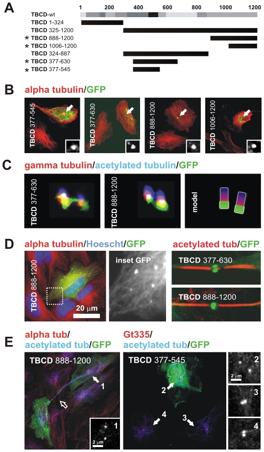
Figure 3. TBCD contains a microtubule-binding region and two centriolar-targeting regions.
(A) Diagram of the full-length TBCD polypeptide and the truncation mutants produced for this study. The degree of evolutionary conservation of the polypeptide is shown in relative shades of grey. (B) Confocal images of HeLa cells transfected with constructs encoding GFP-fusion TBCD truncation mutants and immunostained for tubulin. The constructs shown correspond to those labelled with an asterisk in A. The GFP labelling at the centrosomal region is shown in the inset. (C) High-resolution confocal images of triply labelled centrioles in weakly expressing cells show the co-localization of these two TBCD truncation mutants with γ-tubulin in the proximal region of both centrioles. (D) TBCD contains a microtubule-binding region. GFP–TBCD888–1200-decorated microtubules 48 h after transfection. (Left) TBCD centrosomal-binding truncation mutants were also recruited to the Fleming bodies of cells at the end of mitosis. (E) Phenotypes observed for TBCD centrosomal-binding truncation mutants. Aberrant midbodies with increased lengths and reduced thicknesses were common (right, open arrow). Centriolar separation was increased (>2 µm) in many of the transfected cells (centrosomes 1, 2 versus 3, 4).

