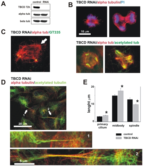
Figure 4. TBCD depletion resulted in microtubule spindle abnormalities and failure of cell abscission.
(A) Western blot confirmation of TBCD silencing 72 h after siRNA treatment. Whole cell lysates (50 µg/lane) were loaded and analysed by immunoblotting with antibodies directed against CoD and the α- and β-tubulins. TBCD depletion did not affect α- or β-tubulin levels. (B) Confocal-microscopic projected images of different mitotic spindle defects observed after TBCD interference in HeLa cell cultures. Mitotic aberrations included abnormally short spindles, abnormal anaphase figures, and multipolar spindle defects, among others. (C) Acentriolar spindle poles were also observed (arrow). (D) TBCD silencing resulted in cell abscission failure. The maintenance of cytoplasmic bridges containing microtubules is shown (1, 2). These abnormally long midbodies are characterized by their content of acetylated microtubules (2). (E) Statistical analysis of the length of the primary cilia (DF = 147; P = 3×10⁻³) and midbodies (DF = 174; P = 0.1) confirmed significant increases in the lengths of these two structures after TBCD depletion. A highly significant reduction in the spindle pole-to-pole distance (DF = 63; P = 5×10⁻⁴) was also detected (*).

