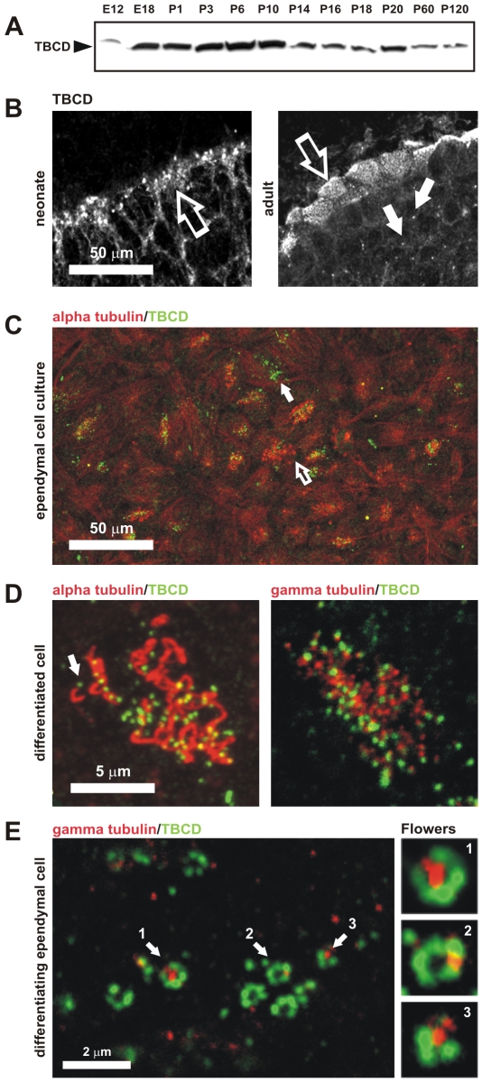
Figure 5. TBCD is required in ciliogenesis.
(A) TBCD is abundantly expressed during neurogenesis. Western blot of total protein extracts (50 µg/lane) from developing and adult mouse brains obtained at the indicated embryonic and postnatal ages. (B) Confocal-microscopic images of cryo-sections of neonatal and adult murine cerebral ependymal epithelium immunostained for TBCD. This cofactor accumulated in clusters localized in the ependymal cell cytoplasm (left, open arrow). In the adult, TBCD localized at the apical borders of the ependymal cells, where the basal bodies are located (right, open arrow). TBCD was also detectable on parenchymal cell centrosomes (solid arrow). (C) A general view of an ependymal cell primary culture in which cilia, immunostained with anti-tubulin antibody, and TBCD are labelled. These culture conditions allow the observation of epithelial cells at various differentiation stages (I–IV, see the text). (Solid arrow) TBCD accumulated in differentiating cells with no cilia, whereas ciliated cells (open arrow) contained fewer TBCD spots. (D) Detail of the apical borders of differentiated ependymal cells where a single TBCD spot was observed at the base of each cilium (arrow). These TBCD spots partially co-localized with the γ-tubulin-labelled basal body. (E) Detail of the cytoplasm of a differentiating ependymal cell (stages I–II), where the assembly of several centriolar rosettes, labelled for TBCD and γ-tubulin, are shown. TBCD accumulated into structures of approximately 0.3 µm in diameter, which created ring-like structures that were often associated with a single γ-tubulin spot of a similar diameter (1, 2, and 3).

