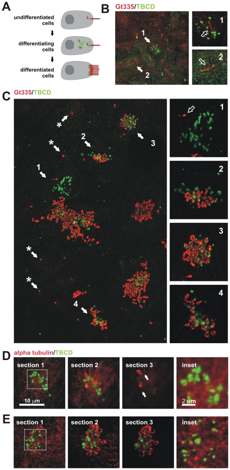
Figure 6. TBCD changes during ciliogenesis in ependymal cells.
(A) Diagram of the location of TBCD accumulation (green) with respect to the centrioles and cilia (red) during ependymal cell differentiation. (B) Short-term cultures display cells at stages I and II, where only a single cilium per cell is observed, presumably the primary cilium. (1) The presence of the primary cilium is often accompanied by the development of small cytoplasmic TBCD clusters containing 1–3 TBCD spots of approximately 0.3 µm. (2) Uncommitted epithelial cells in the culture displayed diffuse TBCD immunostaining. This confocal plane did not coincide with the daughter centriole. (C) General view of a culture immunostained with GT335 and for TBCD. Here, the relationship between TBCD and the assembly of ependymal cilia can be seen. (*) Primary cilia with their corresponding TBCD-labelled centrioles are observed. (1) Differentiating cells (stages I–II) contain TBCD aggregates in their cytoplasm (Figure 5E). (2–4) Cilia assembly occurs after TBCD spots migrate to the apical border of the cells as single spots, presumably accompanying newly developed centrioles. (D-E) A series of confocal sections obtained from the apical borders of the epithelial cells towards the cilia. Cells were immunostained with anti-α-tubulin and anti-TBCD antibodies to show the relationships between this cofactor, the microtubule cytoskeleton, and the cilia. (D) Differentiating cells displaying a few cilia (arrows) show TBCD clusters visible in the cytoplasmic confocal planes (inset). (E) Differentiated cells containing several cilia show smaller TBCD aggregates in the confocal planes, which coincide with the basal bodies of existing cilia (inset).

