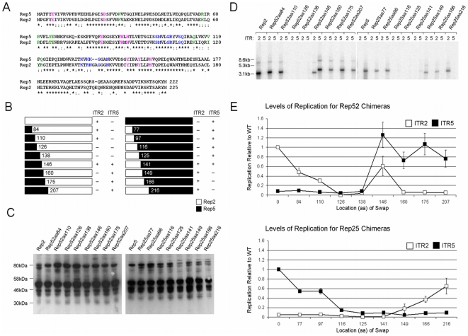
Figure 5. Cloning and characterization of chimeric Reps.
(A) An alignment of the N-termini of Rep2 and Rep5. (*) represents conserved amino acids. (: and.) indicates conservative substitutions. Blue indicates residues implicated in RBE binding interactions. Pink indicates residues which participate in the endonucleolytic active site. Green indicates residues implicated in RBE' binding. (B) Chimeric Reps created and their ability to replicate ITR2 or ITR5 flanked vectors. Numbers indicate the aa position of the switch from one Rep to the other. (+) indicates the presence of replication, (−) indicates the absence. (C) Western blot for expression of the chimeric Reps. (D) Southern blot demonstrating replication of an ITR2 or an ITR5 vector by the chimeric Reps. Note that the ITR5 vector is 500bp larger than the ITR2 vector. (E) Level of replication of the chimeric Reps relative to wt Rep2 or Rep5.

