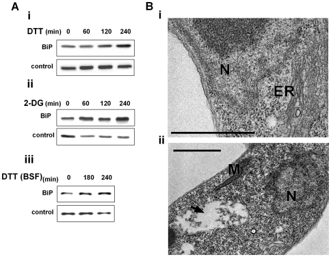
Figure 3. ER stressors induce BiP expression and ER dilation.
A. Western blot analysis of BiP protein. Whole cell extracts (106 cells/lane) were prepared from procyclic (i-ii) or bloodstream trypanosomes (iii) treated with ER stressors for different time points. The extracts were separated on a 10% SDS-polyacrylamide gel and subjected to Western blot analysis using anti-BiP antibodies. hnRNPD0 was used to control loading. i. 4 mM DTT; ii. 20 mM 2-deoxy-D-glucose (2-DG); iii. 4 mM DTT. B. Electron micrographs. i. untreated cells; ii. cells treated with 4 mM DTT for 1 hour. The black arrow indicates the expanded ER. N, nucleus; M, mitochondria; ER, endoplasmic reticulum. Scale bars, 1 µm.

