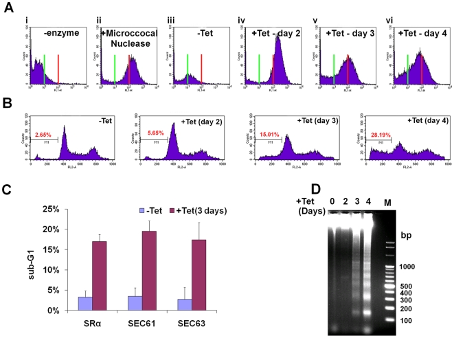
Figure 6. SLS elicits DNA fragmentation.
A. TUNEL assay in SRα silenced cells. TUNEL assay and FACS analysis were performed as described in Materials and Methods. Cells after 3 days of induction without addition of terminal deoxynucleotidyl transferase was used as a negative control. ii. Uninduced cells treated for 10 min with 3000 units/ml of micrococcal nuclease before labeling, used as a positive control. iii. Uninduced cells (-Tet). iv-vi. SRα silenced cells for 2, 3 or 4 days (+Tet), respectively. The green line indicates fluorescence level of cells harboring undamaged DNA. Red line indicates the fluorescence level of cells carrying damaged DNA. B. Analysis of DNA fragmentation by propidium iodide staining and flow cytometry. Uninduced cells (-Tet) or SRα cells silenced for 2, 3 or 4 days (+Tet) were fixed with 100% ethanol for 12 hours at 4°C, washed with PBS and stained for 0.5 hour with 25 µg/ml propidium iodide in the presence of 1 µg/ml RNaseA. DNA content was measured by FACS. The percentage of sub-G1 cells (M1) is indicated. C. Analysis of DNA fragmentation by propidium iodide staining and flow cytometry. The graph represents measurements of sub-G1 population from SRα, SEC61 and SEC63 cells, uninduced (-Tet) or silenced for 3 days(+Tet). The data are derived from three independent experiments and the S.D is indicated. D. DNA laddering during SRα silencing. DNA was isolated from uninduced cells or cells silenced for 2, 3 or 4 days (+Tet) as described in Materials and Methods. DNA (10 µg) was separated on a 1% agarose gel and the gel was stained with ethidium bromide. M - 100bp DNA ladder.

