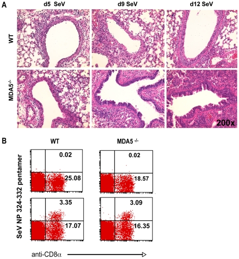
Figure 2. Increased histopathology in MDA5−/− mice.
A) H&E micrographs of lung sections obtained from WT and MDA5−/− mice infected with 400K pfu SeV on d5, d9, d12 PI. B) FACS analysis of lymphocytes derived from the lungs of WT and MDA5−/− mice, uninfected (top panels) and d5 post infected (bottom panels) stained with anti-CD8 and H-2Kb: FAPGNYPAL pentamer.

