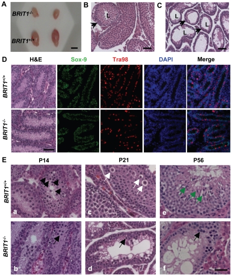
Figure 3. BRIT1-deficient male were infertile and exhibited meiotic defects.
(A) Smaller testes in BRIT1 −/− mice. Shown here are the testes from the indicated mice at P28. Scale bar, 5 mm. (B,C) Thinner seminiferous epithelia in BRIT1 −/− mice. Testes sections from BRIT1 +/+ (B) and BRIT1 −/− (C) mice at P28 were stained with haematoxylin-eosin (H&E). Arrows, the seminiferous tubules; L, lumen of the seminiferous tubules. Scale bar, 50 µm. (D) Mitosis was not defective in BRIT1 −/− spermatogonia and Sertoli cells. Testes at P7 were either stained with H&E or double-stained with anti-Tra98 and anti-Sox9 antibodies using immunofluorescent staining. Scale bar, 50 µm. (E) Meiotic dysregulation in BRIT1 −/− spermatocytes. H&E staining was performed in testis sections from BRIT1 +/+ and BRIT1 −/− littermates at P14, P21, and P56. Black arrows: spermatocytes at leptotene/zygotene (a, b, d, f); white arrows: spermatocytes at diplotene (c); green arrows: elongate spermatids (e). Scale bar, 50 µm.

