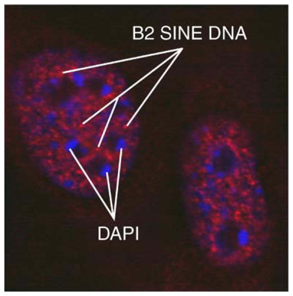
Fig. 3.
FISH microscopy showing distribution of mouse B2 SINE elements. Mouse embryonic fibroblasts were fixed in 1% paraformaldehyde and adhered to slides. Fluorescent oligonucleotides complementary to B2 SINEs hybridized to genomic DNA. There appears to be speckled signal of B2 elements throughout the nucleoplasm, suggesting clusters, but this signal appears not to be preferentially associated with either the nucleoli or the nuclear periphery. Red B2 SINE DNA, blue DAPI stain of AT-rich heterochromatin

