
Figure 1.
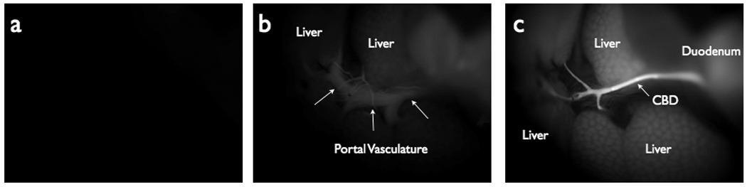
Near Infrared Fluorescent (NIRF) images demonstrating probe accumulation in the biliary system. (A) NIRF image taken prior to probe injection. (B) NIRF image taken at T=1 minutes post-injection, showing probe within hepatic vasculature and initial accumulation in the liver. (C) NIRF image at T=3 minutes post-probe injection, showing high fluorescent signal in the biliary tree and comparatively low signal in the vasculature.