
Table 1.
Parameters Used for Carotid IPH MR Imaging
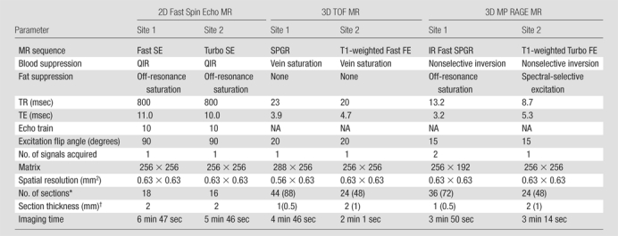
Note.—A field of view of 160 × 160 mm was used for all MR examinations. FE = field echo, IR = inversion recovery, MP = magnetization prepared, NA = not applicable, QIR = quadruple inversion recovery (23), SE = spin echo, SPGR = spoiled gradient recalled echo, TE = echo time, TR = repetition time, 2D = two-dimensional.
*
Values in parentheses are the interpolated numbers of sections after zero-filled Fourier transform in the slab direction.
†
Values in parentheses are the interpolated section thicknesses after zero-filled Fourier transform in the slab direction.