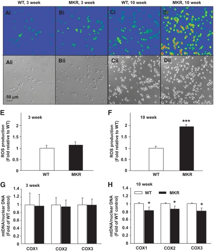
FIG. 6.
Increased pro-oxidant levels and mitochondrial DNA damage in MKR diabetic islet cells. Dispersed islet cells from 3-week-old (A and B) and 10-week-old (C and D) mice were incubated with 10 μmol/l DCF in Krebs-Ringer buffer containing 2.8 mmol/l glucose for 45 min at 37°C. After washing with Krebs-Ringer buffer, cell fluorescence was measured at 480 nm excitation and 510 nm emission using an Olympus fluorescent BX51W1 microscope. A–D: Representative fluorescent (upper panel) and light (lower panel) images of the islet cells. E and F: The average fluorescence intensity was calculated by tracing around each cell and averaging the fluorescence across the entire field of view. (n = 4 with three mice per genotype in each experiment.) mtDNA quantity (G and H) was calculated as the ratio of COX to β-actin DNA levels. (n = 3 with 8–10 mice per genotype in each experiment.) Data are means ± SE. *P < 0.05; ***P < 0.01 compared with age-matched WT. (A high-quality digital representation of this figure is available in the online issue.)

