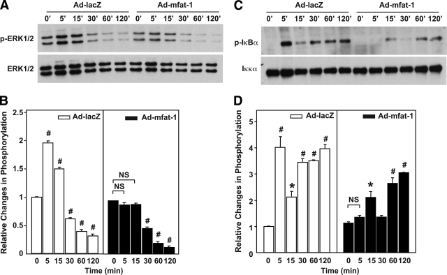
FIG. 3.
Activation of ERK1/2 and NF-κB in INS-1 cells infected with Ad-mfat-1 or Ad-lacZ. INS-1 cells were infected with Ad-mfat-1 or Ad-LacZ for 4 h. Approximately 48 h later, the cells were exposed to IL-1β (5 ng/ml) (A and B) or TNF-α (10 ng/ml) (C and D) for the indicated time. The cells were then extracted for protein for Western blot assays of ERK1/2 and IκB-α phosphorylation. The level of phosphorylation was quantified with Image J software. #P < 0.01, *P < 0.05 when compared with the corresponding control group.

