FIG. 2.
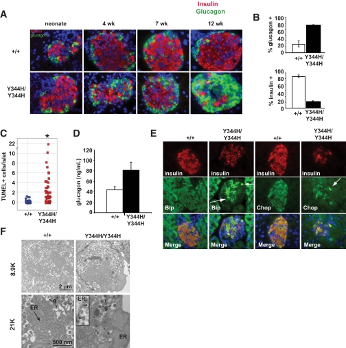
Sec61a1Y344H/Y344H mice lose β-cells by apoptosis and exhibit signs of ER stress. A: Islets from neonate, 4-, 7-, and 12-week-old wild-type (+/+) and mutant (Y344H/Y344H) mice fed HFD stained for insulin (red), glucagon (green), and nuclei (blue) (×600 magnification). B: Quantitation of islet composition from 12- to 15-week-old sex-matched mice fed HFD (n = 3 mice each genotype, 10 islets each). Error bars represent SD. C: Quantitative assessment of TUNEL+ β-cells in islets from three wild-type (+/+) and three mutant (Y344H/Y344H) mice aged 4 weeks and fed HFD for 1 week. Data are expressed as the number of TUNEL+ cells per islet. Statistical assessment was made using a two-tailed Student t test assuming unequal variances. *P < 0.01. D: Glucagon levels measured from 12-week-old sex-matched mice on HFD (n = 5 MUT; n = 6 wild type). Values shown are means ± SEM. E: Islets from 4-week-old wild-type (+/+) and mutant (Y344H/Y344H) mice fed HFD for 1 week stained for markers of ER stress (Bip [left two columns; green] and Chop [right two columns; green]), insulin (red), and nuclei (blue) (×600 magnification). Arrows highlight those β-cells with high expression of either Bip or Chop. F: Electron microscopy of β-cells from islets isolated from a wild-type (+/+) and mutant (Y344H/Y344H) mice aged 6 months. Note normal secretory granule (g) morphology, but marked ER dilation. Inset: blowup of the ER membrane demonstrating ribosomes (arrowheads) free in the cytosol and bound to the ER membrane. (A high-quality digital representation of this figure is available in the online issue.)

