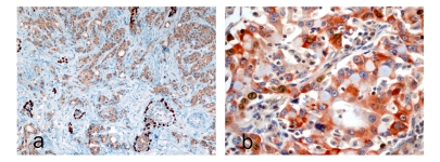
Figure 1.
a. Human prostate core biopsy with double immunohistochemical staining for high molecular weight cytokeratin (K903) and AMCAR (alpha-methyl-CoA-racemase). The dark brown stain (K903) highlights the basal epithelial cells and the light brown cytoplasmic stain AMCAR in prostate cancer cells including dysplastic cells in high-grade prostatic intraepithelial neoplasia (HGPIN). The differential localization and distribution are useful in confirming areas of invasive carcinoma (×40) in addition to conventional criteria for malignancy. b. Bcl-2 (anti-apoptotic protein) and Ki-67 in human lung carcinoma (courtesy Epitomics,Inc). This also highlights differential localization of the two proteins; Bcl-2 to cytoplasm and Ki-67 nuclear and also suggests that Ki-67 staining cells are different from Bcl-2 staining cells and the transcription cycle of the proteins.

