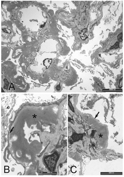
Figure 2.
Transmission electron microscopy. A. Prominent deposits associated with the mesangium and GBM. B. Higher power view showing a deposit (*) in the subendothelial space adjacent the internal aspect of the GBM (arrow). C. Deposit (*) adjacent the mesangium with basement membrane (arrow). Note that the deposits are more electron dense than basement membrane

