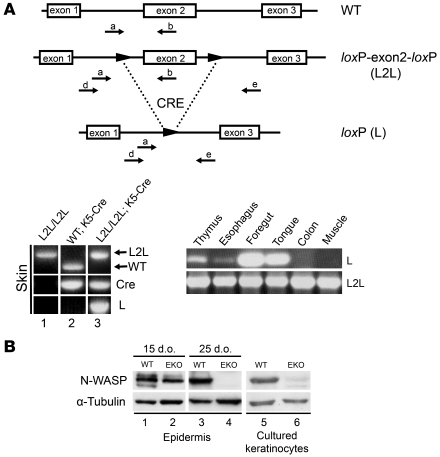
Figure 1. Epidermis-specific KO of N-WASP.
(A) Top: PCR strategy to detect exon 2 deletion of N-Wasp. Primers used to determine presence (a, b) or absence (d, e) of exon 2. Bottom left: PCR amplification of tail DNA from 4-month-old N-WaspL2L/L2L (L2L/L2L) mice with and without the K5-Cre transgene and a WT mouse with the K5-Cre transgene. Amplification of WT and L2L/L2L DNA with primers a and b resulted in a 350-bp band (lane 2 WT) and a 550-bp band (lane 1 L2L), respectively. Amplification of WT DNA with primers d and e failed to yield a product (lane 2 L). Amplification with primers d and e resulted in a 1.5-kb band, thus identifying the excision of exon 2 in L2L/L2L; K5-Cre mice (lane 3 L). The presence or absence of Cre is noted by separate PCR amplification in the middle row. Bottom right: Gel electrophoresis of DNA from different tissues of a 4-month-old N-WaspL2L/L2L/K5-Cre mouse following PCR with primer sets to determine the presence (L2L, primers a and b) or absence (L, primers d and e) of exon 2. Exon 2 excision was observed in thymus, esophagus, foregut, and tongue. (B) Western blot analysis of N-WASP expression in epidermis (lanes 1–4) and cultured keratinocytes (lanes 5 and 6) isolated from L2L (referred to as WT) and N-WaspL2L/L2L/K5-Cre (L2L; K5-Cre – i.e., EKO) mice. Epidermis was isolated from 15- and 25-day-old mice, and primary keratinocytes were isolated from 3-day-old mice and maintained in culture for 5 days. N-WASP expression was undetectable in EKO mice.

