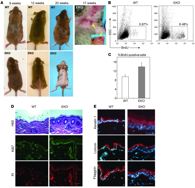
Figure 2. Hair loss, ulceration, and epidermal hyperproliferation in N-WASP EKO mice.
(A) WT and EKO littermate mice 9, 12, and 20 weeks old. Ulcers (arrows) on the chest and face of a 17-week-old EKO mouse are also shown. (B) Representative FACS analysis of primary keratinocytes. Keratinocytes were isolated from mice 2 hours after BrdU injection and analyzed by flow cytometry. Selected (gated) events represent the total percentage of BrdU+ keratinocytes in the representative experiment; average BrdU+ in EKO mice was 5.1% (n = 11) vs. WT 1.0% (n = 10) (P = 0.004). (C) Results of proliferation analysis of primary keratinocytes in culture. Cultured keratinocytes were labeled with BrdU for 30 minutes, fixed, stained with anti-BrdU antibody, and analyzed by flow cytometry. The results of 1 representative experiment (repeated 3 times) are shown; each bar represents mean value of 6 samples ± SD. (D) H&E-stained paraffin skin sections of WT and EKO mice demonstrate an expansion/hyperproliferation of HFs and interfollicular epidermis (upper panel, arrows). Frozen skin sections were stained with the antibody against the proliferation marker Ki-67 (green), and nuclei were stained with propidium iodide (PI, red). The increased epidermal proliferation was associated with an increase in Ki-67+ nuclei. Scale bar: 100 mm. (E) Normal distribution of early (keratin 1) and late (loricrin, filaggrin) markers of differentiation in hyperproliferative N-WASP–deficient epidermis. Frozen sections stained with antibodies against keratin 1, loricrin, and filaggrin (red). Nuclei were counterstained with Hoechst (blue). Scale bar: 40 mm.

