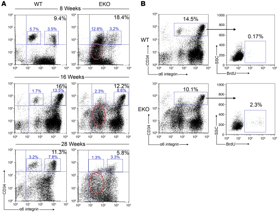
Figure 5. N-WASP EKO mice exhibit a progressive decline in HF stem cells.
(A) Representative FACS analysis on primary keratinocytes isolated at 8, 16, and 28 weeks of age revealed an initial increase in CD34+/α6+ progenitor cells in N-WASP EKO mice. This population diminished over time and was dramatically decreased at 28 weeks. Additionally, there was an early appearance and persistence of a CD34lo/–/α6lo population (red circles) corresponding to an abnormal expansion of ORS keratinocytes in N-WASP EKO mice. Selected (gated) events in each plot represent the percentage of α6lo (left boxes) and α6hi (right boxes) cells in the total CD34+/α6+ population. (B) FACS analysis of BrdU incorporation by keratinocytes gated for the stem cell population revealed increased proliferation specifically within this population in N-WASP EKO mice. Gated events in the left panels represent the percentage of CD34+/α6+ keratinocytes. Gated events in the right panels indicate the percentage of BrdU+ keratinocytes within the selected CD34+/α6+ population.

