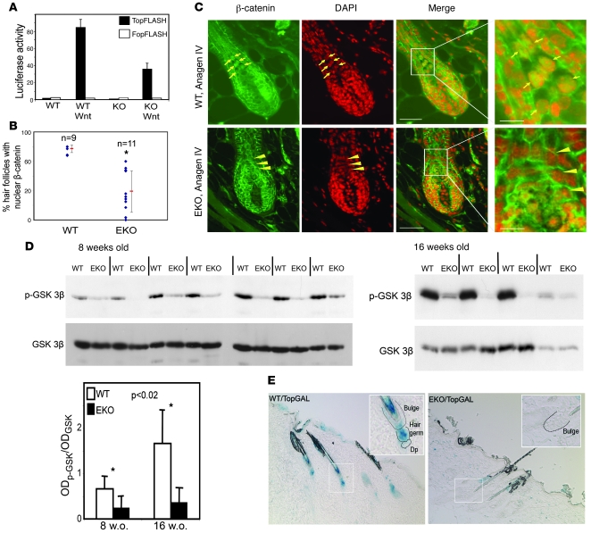
Figure 6. Defective Wnt signaling in N-WASP–deficient epidermis.
(A) Decrease in Wnt-dependent transcription in N-WASP KO fibroblasts. WT and N-WASP KO fibroblasts or cells expressing a Wnt-responsive (TopFLASH) or a mutant (FopFLASH) reporter construct. Bar graph represents reporter activity upon Wnt stimulation (normalized). Error bars represent SD. Representative results of 1 of 3 experiments are shown. (B and C) Defect in nuclear localization of β-catenin in N-WASP–deficient HF keratinocytes. Anagen IV follicles in 8-week-old WT (C, upper panels) and N-WASP EKO (C, lower panels) mice. Green indicates β-catenin antibody staining; red indicates nuclei stained with DAPI; arrows indicate β-catenin–positive nuclei in precortical keratinocytes in WT follicles; arrowheads indicate nuclei devoid of β-catenin in precortical cells of N-WASP–deficient follicles. Scale bar: 40 mm (inset bar, 12 mm). Scatter plot (B) represents the percentage of anagen IV follicles with nuclear β-catenin in the subcortex in WT and N-WASP EKO mice. Each dot represents 1 mouse. Vertical lines indicate the mean (middle dash) ± SD. *P < 0.001. (D) Decrease in serine 9–phospho–GSK-3β in N-WASP EKO skin. Each lane on the Western blot represents 1 mouse. Bar graph depicts the OD of the phospho–GSK-3β band (upper panel) normalized to the OD of the total GSK-3β band (lower panel). Error bars represent SD. (E) Decreased Wnt-dependent transcription in N-WASP EKO HFs. Skin from non-depilated 8-week-old WT/TopGAL and EKO/TopGAL mice was processed for X-gal staining. Blue color corresponds to active Wnt-dependent transcription. Original magnification, ×10 (inset, ×20).

