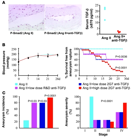
Figure 1. Neutralization of TGF-β activity induces a highly permissive state for Ang II–induced aortic aneurysm formation and complications.
(A) Phosphorylation of Smad-2 (nuclear red staining) is abrogated in the vascular media and adventitia of aortas recovered 3 days after intraperitoneal administration of 500 μg of anti–TGF-β antibody (2G7 clone). Note that at this time point, the media and adventitia are infiltrated by macrophages (see Figure 3E). Original magnification, ×200 for Ang II and ×100 for Ang II + anti–TGF-β. Right panel: Serum TGF-β is barely detectable in mice 3 days after 500 μg of anti–TGF-β antibody compared with nontreated animals. Results are representative of 2 different experiments. (B) Neutralization of TGF-β activity did not alter blood pressure levels (left panel). Right panel: Effects of low-dose anti–TGF-β antibody (R&D Systems, n = 5; or 2G7 clone, n = 5) or high-dose 2G7 clone (n = 26) on survival from ruptured aortic aneurysm, compared with mice that received Ang II in the absence of TGF-β neutralization (n = 29). (C) Quantification of aneurysm incidence and severity (increasing severity from stage I to stage IV as described in ref. 52) in all groups of mice. Stage IV was attributed to ruptured aneurysms.

