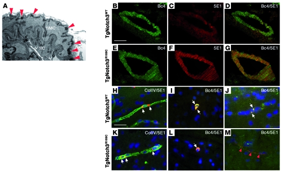
Figure 3. Notch3ECD aggregates and GOM deposits in brain arteries and capillaries of TgNotch3R169C mice.
(A) Electron micrograph of a pial artery from a 5-month-old TgNotch3R169C mouse demonstrating abundant GOM deposits (red arrowheads) within the basement membrane of smooth muscle cells. (B–G) Brain arteries from 2-month-old TgNotch3R169C and TgNotch3WT mice were stained with Notch3 antibodies specific to the intracellular (Bc4, green) or extracellular (5E1, red) domain. Microscopic aggregates of Notch3ECD are shown in mutant artery. (H, I, K, and L) Brain sections of 2-month-old TgNotch3WT and TgNotch3R169C were double labeled with antibodies to Notch3 extracellular domain (5E1, red) and collagen IV (ColIV, green) (H and K) or double labeled with Bc4 (green) and 5E1 (red) Notch3 antibodies (I and L). Nuclei were stained by DAPI (blue). Perinuclear inclusions, strongly labeled by Notch3 intracellular and extracellular antibodies, were seen in wild-type and mutant capillaries (white arrows). (J and M) Brain sections of 12-month-old TgNotch3WT (J) and TgNotch3R169C (M) were double labeled with Bc4 (green) and 5E1 (red) Notch3 antibodies. Shown are dot-like Notch3ECD aggregates unstained by the Bc4 antibody in the mutant capillary (red arrowheads), while wild-type capillary exhibited discrete perinuclear inclusions labeled by Notch3 intracellular and extracellular antibodies (white arrows). Scale bars: 1 μm (A), 50 μm (B–G) and 30 μm (H–M).

