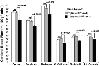
Figure 5. Decreased resting CBF in aged TgNotch3R169C mice.
Quantitative measurement of CBF through the neocortex, forebrain (including the striatum, pallidum, and amygdala), thalamus, corpus callosum, fimbria of the hippocampus, and internal capsula in 18-month-old TgNotch3R169C compared with TgNotch3WT and nontransgenic mice showed diffuse cerebral hypoperfusion in mutant mice. Each of these large areas included 8–10 gray matter regions or 4–5 white matter regions.

