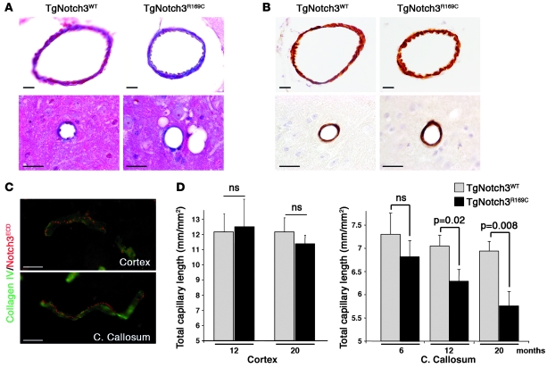
Figure 6. Arterial structure is preserved, but capillary density is progressively reduced in the white matter of TgNotch3R169C mice.
(A) Masson’s trichrome staining showing intact pial artery (upper panels) and small artery (lower panels) of 20-month-old TgNotch3R169C mice. (B) Labeling for smooth muscle myosin heavy chain revealing a continuous rim of smooth muscle cells in pial and small artery of 20-month-old TgNotch3R169C mice. (C) Double labeling for Notch3ECD (5E1, red) and collagen IV (green) of capillaries in the cortex and the corpus callosum of 12-month-old TgNotch3R169C mice showed robust aggregation of Notch3ECD in both capillaries. (D) Mean total capillary length (mm of CD31+ structures per mm2) in the cortex of 12- and 20-month-old TgNotch3R169C mice and the corpus callosum of 6-, 12-, and 20-month-old TgNotch3R169C mice compared with age-matched TgNotch3WT mice showed progressive age-related reduction in capillary density in the white matter of mutant mice (n = 4–5 mice per group). Scale bars: 25 μm.

