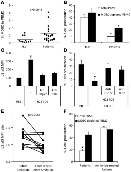
Figure 8. Exosomes produced by human cancer cell lines or metastatic cancer patients dictate Stat3 activation in MDSCs and their immunosuppressive function through TLR2 and Hsp72.
(A) The frequency of MDSCs, defined as HLA-DR CD33+ cells, is shown in the PBMCs of healthy volunteers (H.V.) (n = 11) and metastatic cancer patients (n = 18). Each plot is an individual measure, and the horizontal bar is the mean. (B) Immunosuppressive function of MDSCs from peripheral blood of healthy volunteers and metastatic cancer patients on stimulated T cell proliferation. T cell stimulation was induced by a mixture of anti-CD2, anti-CD3, and anti-CD28 beads (n = 10). (C) PBMCs from healthy volunteers were cultured for 24 hours in medium alone or medium containing TDEs from H23 cells with or without blocking TLR2 Abs or anti-Hsp72 polyclonal Abs (pAbs). pStat3 was determined by flow cytometry on MDSC gated cells (n = 10). (D) Immunosuppressive function of MDSCs from blood of healthy volunteers either untreated or treated with TDEs from H23 cells alone or with blocking TLR2 Abs or anti-Hsp72 pAbs (n = 8). (E) PBMCs from metastatic cancer patients were incubated overnight in serum-free medium supplemented with autologous serum or PBS. pStat3 expression in gated MDSC was determined by flow cytometry. pStat3 MFI ratio between PBS and serum condition was represented. The same patients were sampled before and after 3 weeks of amiloride treatment (n = 11). (F) Immunosuppressive function of MDSCs prepared from peripheral blood of metastatic cancer patients, treated with amiloride or not treated, on T cell proliferation stimulated as in B. *P < 0.05. Error bars represent mean + SD.

