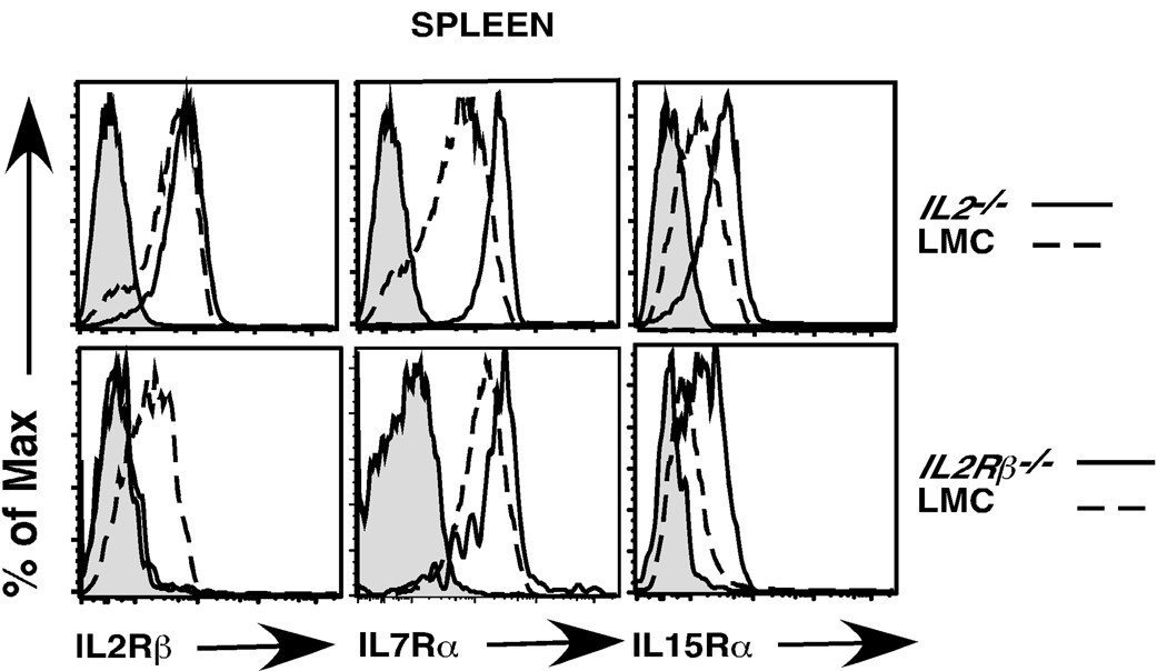
Fig. 6. Expression of IL2Rβ, IL7Rα and IL15Rα on CD4+Foxp3+ Tregs from IL2−/− and IL2Rβ−/− mice compared to wild type littermate controls.
Splenocytes were isolated from 4–5 week old LMC, IL2−/− and IL2Rβ−/− mice and stained with antibodies for CD4, CD8, and Foxp3 to identify splenic Tregs. Shown are CD4+Foxp3+ gated cells stained for IL2Rβ (left panels), IL7Rα (middle panels) and IL15Rα (right panels). Gray histograms represent staining of CD4+Foxp3+ cells with isotype control antibody. Solid lines represent histograms of CD4+Foxp3+ T cells from IL2−/− (top panel) or IL2Rβ−/− (bottom panel) mice; broken lines represent staining of CD4+Foxp3+ Tregs from WT LMC mice. A representative example of 3 independent experiments is depicted (n= 6 IL2−/− and 3 IL2Rβ−/− mice).

