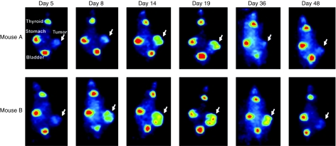
Figure 4.
In vivo monitoring of MV-NIS infection over time. Two mice harboring LNCaP xenografts received a single IV injection of 1.5 × 10⁶ TCID50 measles virus–sodium iodide symporter (MV-NIS) and viral biodistribution and kinetics were monitored by 123I imaging. Initially, MV-NIS carried by the blood supply into the xenograft could be detected in small, localized sites inside the tumor. By day 14, the replicating virus had spread extensively throughout the tumor mass and strong, persistent infection could be detected for as long as 36 days. MV-NIS replication inside the tumor of mouse A resulted in significant partial regression of the growth that relapsed when the virus was cleared (negative uptake signal). Mouse B exhibited tumor growth arrest (stable disease) due to persistent MV-NIS infection. The virus was ultimately cleared from the mouse and tumor growth was reinstated.

