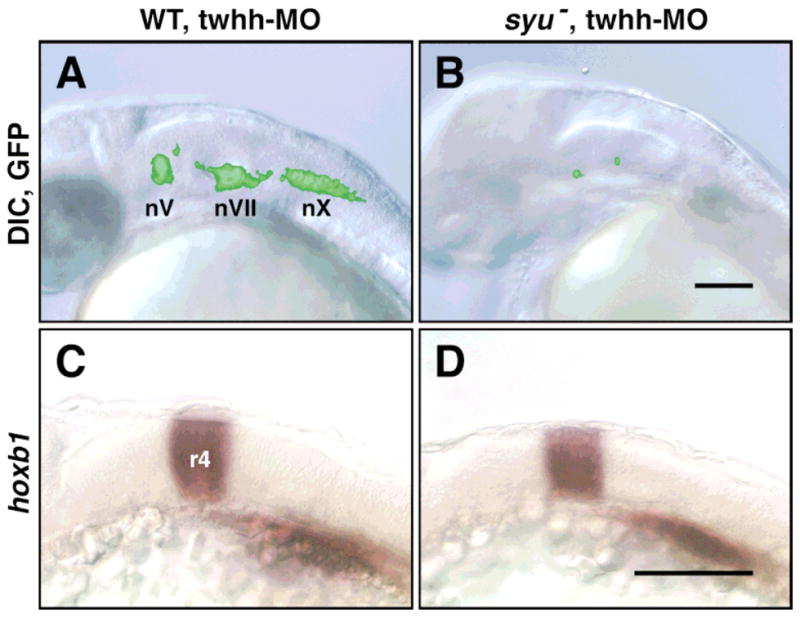
FIG. 2.

Hindbrain development and patterning are not affected in “double mutants.” All panels show side views of the hindbrain with anterior to the left. (A, B) Live, 30 HPF embryos were embedded in 3% methylcellulose and photographed using DIC optics and GFP epifluorescence. The fluorescent images of the GFP-expressing cells were subsequently superimposed on the DIC images using Photoshop software. In a twhh MO-injected wild-type embryo (A), the GFP-expressing BMNs (nV, nVII, nX) are found in normal numbers at characteristic locations. In contrast, in a twhh MO-injected syu “double mutant” (B), very few GFP-expressing cells are found in an otherwise healthy hindbrain. (C, D) Twhh MO-injected embryos were examined under epifluorescence at 23 HPF to select wild-type (n = 5) and “double mutant” (n = 3) embryos, which were processed for hoxb1 in situ hybridization. Hoxb1 is expressed normally in rhombomere 4 in twhh MO-injected wild-type (C) and syu mutant embryos (D). Scale bars = 100 μm.
