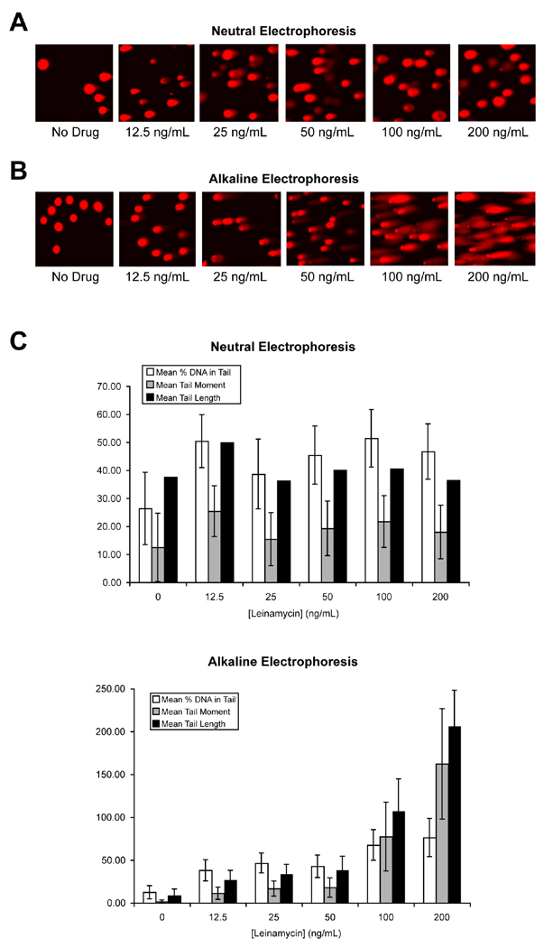
Figure 4.
Level of leinamycin-induced DNA strand breaks breaks in MiaPaCa cell as determined by a neutral Comet assay. MiaPaCa cells were treated with 0, 12.5, 25, 50, 100, or 200 ng/mL leinamycin for 2 hours, and the lysis of the harvested cells was done under neutral conditions. The electrophoresis was done under either neutral (A) or alkaline (B) condition. C, Mean tail moment, mean tail length, and mean percentage of tail DNA in MiaPaCa cells treated with 0, 12.5, 25, 50, 100, or 200 ng/mL leinamycin for 2 hours and analyzed by a neutral Comet assay using electrophoresis under either neutral or alkaline condition. The number of cells scored in each treatment was 30–60. Error bars denote SEM.

