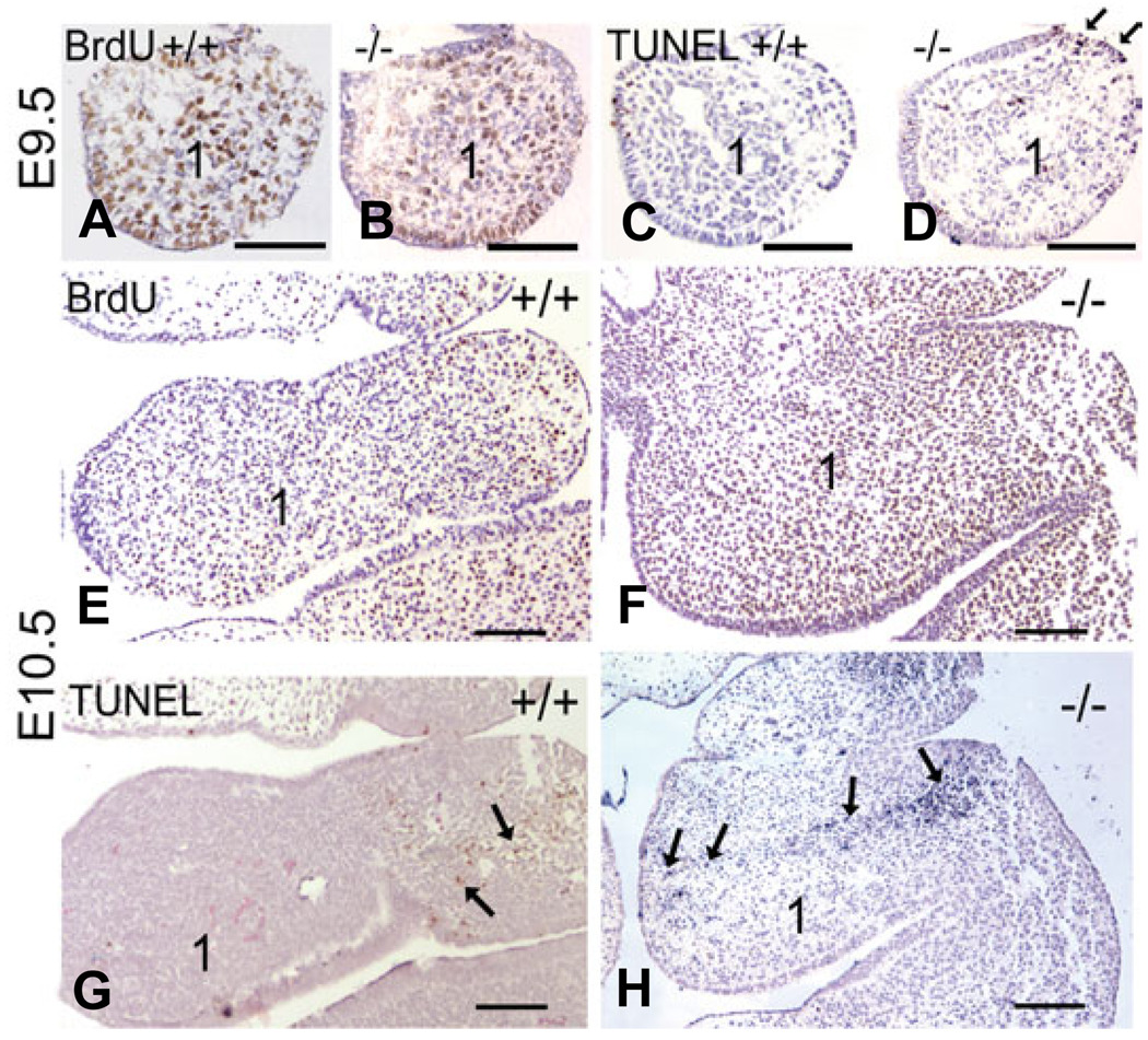
Fig. 6. Comparison of proliferation and cell death in Ednra−/− embryos.
Transverse sections through the mandibular arch one and arch two of wild type (A,C,E,G) and Ednra−/− (B,D,F,H) following BrdU (A,B,E,G) or TUNEL (C,D,F,H) analysis. (A,B) BrdU labeling in the mandibular arch (1) appears similar between wild type (A) and Ednra−/− (B) embryos. (C,D) In consecutive sections to those in (A,B), few TUNEL-positive cells are observed in the mesenchyme of E9.5 wild type embryos (C), though numerous labeled nuclei (red) are observed in the mesenchyme of Ednra−/− embryos (D). There is also a local increase in TUNEL-positive cells in the ectoderm of the proximal arch (arrows in D). (E,F) At E10.5, BrdU staining appears similar between wild type (E) and Ednra−/− (F) embryos in the first arch. (G,H) In consecutive sections to those in (E,F), TUNEL-positive cells (red) in wild type embryos are confined to the proximal aspects of arches one and two (arrows in G). In Ednra−/− embryos, TUNEL-positive cells (blue) extend from this proximal region into the distal arch as denoted by arrows (H; the difference in color between labeled cells in G and H is due to different chromagens. This does not affect the outcome or sensitivity of the experiments). The magnification bar in each panel represents 100 µm.

