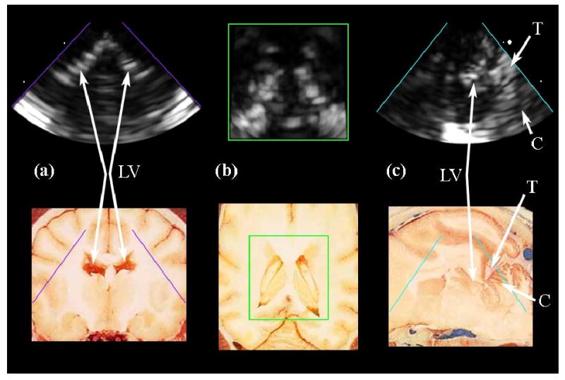
Fig. 10.

(a,b,c) Intracranial RT3D echo images in coronal, axial, and sagittal planes, respectively, compared to corresponding anatomical images (Reproduced with permission from Fletcher et al., University of Minnesota College of Veterinary Medicine). The lateral ventricles (LV) are clearly seen in (a) and (b). The tentorium (T) and cerebellum (C), as well as a posterior horn of the lateral ventricle, are visible in (c).
