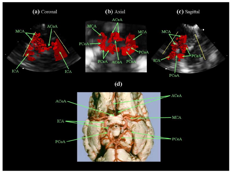
Fig. 11.

(a,b,c) Intracranial RT3D color Doppler images in coronal, axial, and sagittal planes, respectively (note: the color Doppler look-up table was modified, eliminating directional flow information). (d) Latex-injected sheep brain vasculature for anatomical reference (Reproduced with permission from R.R. Miselis, University of Pennsylvania School of Veterinary Medicine). The internal carotid arteries (ICA), left middle cerebral artery (MCA), and anterior communicating artery (ACoA) are visible in (a). The Circle of Willis is shown in (b), with the anterior cerebral arteries (ACeA), posterior cerebral arteries (PCeA) and posterior communicating arteries (PCoA) also indicated.
