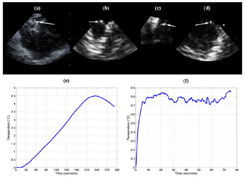
Fig. 12.

(a) AcuNav image of thermocouple (arrow) placed at 1 cm depth in cerebrum. (b,c,d) RT3D coronal, axial, and sagittal images, respectively, of inserted thermocouple (arrows), acquired with dual-mode catheter. (e) Temperature rise achieved using the AcuNav (transducer self-heating and conduction). (f) Temperature rise achieved by driving the dual-mode catheter's linear array elements as a single channel (unfocused) with an RF power amplifier.
