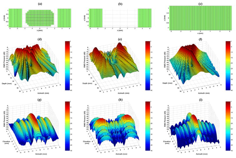
Fig. 3.

Field II simulation apertures and beam plots. (a,b,c) Active apertures for each case: the entire integrated array with 2 cm focus, the integrated catheter's linear arrays with unfocused transmit, and the AcuNav with 2 cm focus, respectively. (d,e,f) Relative pressure amplitude in the zero-elevation plane for each case, respectively. (g,h,i) Relative pressure amplitude at a depth of 2 cm for each case, respectively.
