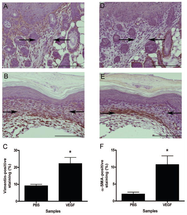
Figure 4. Enhancement of fibroblast and myofibroblast numbers in VEGF-treated fetal wounds.
Immunohistochemical staining for vimentin and α-SMA were used to detect fibroblasts and myofibroblasts, respectively, in E15 fetal wounds at 7 days post-wounding. Graphs demonstrating the density of staining as determined by image analysis and images of representative wounds are shown demonstrating an increase in both vimentin-positive fibroblasts (a–c, left panels) and α-SMA-positive myofibroblasts (d–f, right panels) in wounds injected with 0.1 μg rmVEGF164 (b and e) compared to control wounds injected with PBS (a and d). Arrows are used to indicate the wound bed/scar margins (scale bar = 100 μm). The bars and lines represent means +/− S.E.M.; *p<0.01, unpaired t-test; n = 5 for PBS and n = 4 for VEGF.

