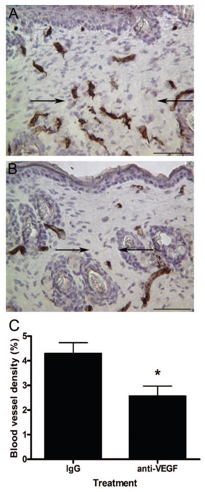
Figure 5. VEGF blockade and wound vascularity.

Immunohistochemical staining for PECAM was used to identify blood vessels in wounds from mice injected with goat IgG (a) or neutralizing anti-VEGF (b) antibodies 14 days post-wounding. The margins of the wound bed/scar are marked with arrows (scale bar = 100 μm). Blood vessel densities were determined and are represented graphically (c). The bars and lines represent means +/− S.E.M.; *p<0.0199, unpaired t-test; n = 6 for IgG and n = 5 for anti-VEGF for each dataset.
