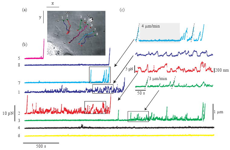
Fig. 2.

Multiplexed force measurements. (a) (Media 1) Eight apCAM-coated beads are held on the membrane of an Aplysia growth cone with holographic optical tweezers. Bead trajectories are superimposed (points represent bead position every 50 s). Movie: sped up 120 times, field of view 75 × 65 μm, arrows indicate forces generated by the cell. (b) Magnitude of bead displacements and measured forces generated by the cells. (c) Close up of the forces for four of the beads.
