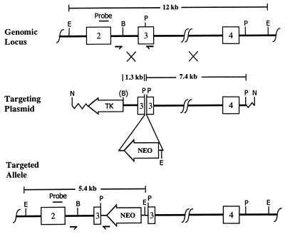
Figure 1.
Targeted disruption of the ERβ gene. (Top) The unmodified genomic locus showing exons 2–4. The probe used for Southern blots is labeled above exon 2. The small arrows indicate the positions of primers. Restriction enzyme sites are EcoRI, BglII, and PstI, designated by E, B, and P. (Middle) The targeting plasmid with the left (1.3-kb) and right (7.4-kb) regions of homology. The Neo insertion site is indicated. NotI (N) was used to linearize the plasmid. Homologous recombination is indicated by the large Xs. (B) shows the BglII lost in the construct. (Bottom) The targeted allele with the Neo gene inserted into exon 3. Note the additional EcoRI site introduced during the targeting. Mouse genomic DNA is shown as a thick line, the inserted Neo sequences as a thinner line, and the plasmid vector as a zig-zag line.

