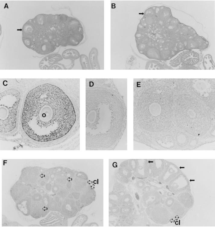
Figure 3.
Histology and ERβ immunocytochemistry in wild-type and ERβ −/− ovary. Hematoxylin-stained sections of adult wild-type (A) and ERβ −/− (B) ovary at ×40 magnification; the arrows indicate mature follicles. (C) Immunocytochemistry of wild-type ovary with an antiserum against ERβ; o, oocyte within the follicle. (D) Immunocytochemistry of wild-type ovary with the antiserum preabsorbed with immunogenic ERβ peptide. (E) Immunocytochemistry of adult ERβ −/− ovary with the ERβ antiserum. Histologic sections of a representative ovary from an immature wild-type female (F) and from an immature ERβ −/− female (G), both after superovulation; several unruptured preovulatory follicles in G are indicated by solid arrows. Corpora lutea (cl) are indicated in F and G by open arrows.

