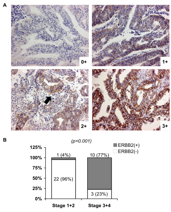
Figure 3.
ERBB2 protein expression in early and late EECs. (A) Representative immunohistochemical (IHC) staining of ERBB2 protein in primary EEC tissues. Staining results were graded as 0+: undetectable staining in <10% of the tumor cells; 2+: weak to moderate complete membrane staining (indicated by an arrow) in <10% of the tumor cells; 3+: strong complete membrane staining observed in <10% of the tumor cells. EEC cases were categorized as ERBB2-negative (scores 0 and 1+) or positive (scores 2+ and 3+). (B) A histogram summarizing the IHC results on 36 primary EEC tissues stained for ERBB2. A chi square test P value is shown. Case numbers and percentages are also indicated.

