
Figure 7. Effects of HDI on ERα and ERβ expression and activity in MCF-7 cells.
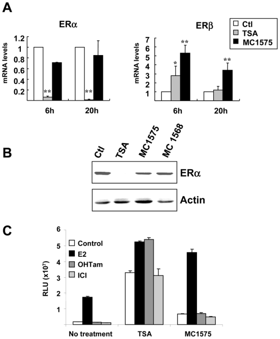
A- MCF-7 cells were treated for 6h or 20 h with TSA (1.7 μM), MC1575 (20 μM) or vehicle alone (Control - Ctl) and mRNA levels for ERα and ERβ were quantified using RT-qPCR. Results are expressed relative to the TBP housekeeping gene and to the mRNA levels measured for the control cells used as reference. Results represent mean and s.d of at least 4 independent cell cultures. Raw data were used for statistical analysis. * p < 0.05, ** p ≤ 0.01 as compared to control cells.
B- MCF-7 cells were treated for 20 h with TSA (1.7 μM), MC1575 (20 μM), MC1568 (20 μM) or vehicle alone (Control -Ctl) and ERα protein levels were analyzed by western immunoblotting. Actin was used as a loading control.
C- MELN cells were treated for 20h with control vehicle (Control), or 17β-estradiol (E2; 10−8M), OHTam (10−8M) or ICI (10−8M) in the absence or presence of TSA (1.7 μM) or MC1575 (20 μM) and luciferase activity was quantified. Results are expressed as relative luciferase units (RLU) and represent mean and s.d. of triplicate wells. These results are representative of 3 independent experiments.