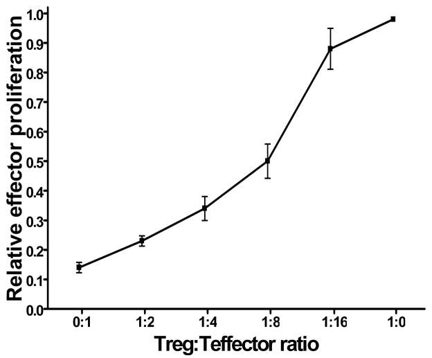
Figure 3. CD4+CD25+ T cells isolated from prostate tumor draining lymph nodes are functionally suppressive.
T cells from tumor-draining lymph nodes isolated from young (≤ 8 weeks old, n = 3) TRAMP mice were purified into CD4+CD25− (responder) and CD4+CD25+ populations by magnetic separation. 5×10⁴ CD4+CD25+ T cells were cocultured for 72 hours with 5×10⁴ allogenic T cell-depleted splenocytes from C3H mice in complete RPMI supplemented with 1 ug/ml activating anti-mouse CD3 antibody, either alone or with autologous CD4+CD25– responder T cells in 1:2, 1:4, 1:8 and 1:16 ratios. As positive controls for maximal proliferation, 5×10⁴ CD4+CD25− responder T cells were cocultured with 5×10⁴ allogenic T cell-depleted splenocytes from C3H mice in complete RPMI supplemented with 1 ug/ml activating anti-mouse CD3 antibody. 3H-thymidine (1 μg/well) was added in the last 8 hours of culture. Responder cell proliferation was measured by 3H-thymidine incorporation. The relative proliferation index of responder cells for each mouse at each Treg:Tresponder ratio was calculated by dividing the mean Tresponder proliferation at each ratio by the maximal proliferation (Tresponders cultured in the absence of Treg) of Tresponders in that animal.

