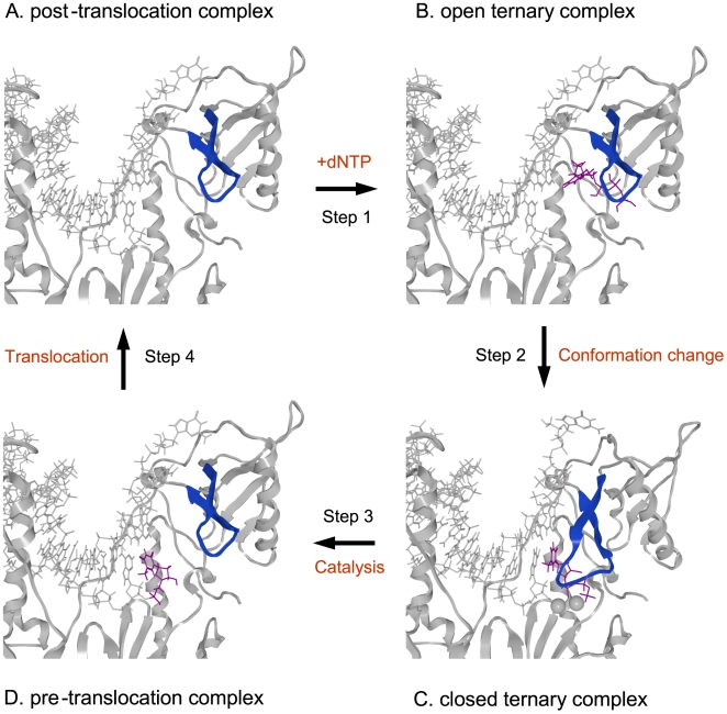
Figure 3. Structural models of the HIV-1 RT p66 subunit in a DNA polymerization cycle.
The 3-D models of the 93JP-NH1 p66-template-primer ternary complex of the fingers-open configuration at post-translocation (A), fingers-open configuration at the stage of dTTP binding (B), fingers-closed configuration after fingers-rotation (C), and fingers-open configuration at pre-translocation stage (D). The models were constructed by homology modeling and docking simulation techniques using two crystal structures [1], [14] of the HIV-1 RTs as modeling templates (see Materials and Methods). Catalytic clefts composed of fingers, palm, and thumb subdomains are shown. dTTP, magenta sticks; p66 main chain, grey ribbon; Mg2+ ion, gray spheres; template-primer, grey sticks; β3-β4 loop of the fingers subdomain, blue ribbon.

